
内容精选
Content Selection
中国佳稿
Articles from China
[Heart] A 40-year-old man with bicuspid aortic valve and huge LV [40岁男性患二叶式主动脉瓣及巨大左心室]
- 分享:
Authors
Yongshi Wang, Lili Dong, Xianhong Shu
Clinical introduction
A 40-year-old man presented to our institute with progressive exertional dyspnoea for 1 year. Bicuspid aortic valve (BAV) with mild-to-moderate aortic regurgitation (AR) was diagnosed in his childhood by echocardiography, but he declined follow-up afterwards. Physical examination revealed a grade 3/6 ‘to-and-fro’ murmur at the third left intercostal space. Chest radiograph showed an extremely enlarged cardiac silhouette (figure 1A), and ECG displayed complete left bundle branch block with frequent premature ventricular contractions. Transthoracic echocardiography demonstrated an extremely enlarged LV with end diastolic volume of 472 mL and EF of 20% (see figure 1B and C).
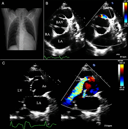
Figure 1 Chest radiograph (A), short axis (B) and long axis (C) transthoracic imaging of the aortic valve. BAV, bicuspid aortic valve. LA, left atrium; RA, right atrium; Ao, aorta.
Question
Based on these imaging and clinical findings, what is the most likely cause of the patient's AR?
A. Aortic dissection
B. Aortico-left ventricular tunnel
C. Avulsion of anterior cusp from aortic annulus
D. Ruptured paravalvular abscess
E. Ruptured sinus of Valsalva aneurysm
Answer: B
The correct answer is aortico-left ventricular tunnel (ALVT). Echocardiography (figure 1B, C) and multislice CT angiography (figure 2A, B) revealed a tunnel-like structure with significant anterograde systolic and retrograde diastolic flow originating from the aortic root just above the level of the dilated aortic sinus and winding leftwards down into the LV, which was the signature feature of ALVT. The final diagnosis of type II ALVT, characterised as extracardiac aortic wall aneurysm of the tunnel with or without valvular distortion, was confirmed by surgical inspection (figure 2C), and the patient performed well after surgical closure of the tunnel.
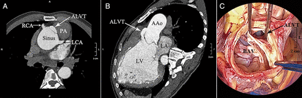
Figure 2 Multislice CT angiography demonstrated a tunnel-like structure originating from the aortic root just above the level of the aortic sinus and winding leftwards down into the LV (A and B). The diagnosis of type II aortico-left ventricular tunnel (ALVT) was confirmed by surgical inspection (C). BAV, bicuspid aortic valve. LA, left atrium; LCA, left coronary artery; RCA, right coronary artery; PA, pulmonary artery. AAo; ascending aorta.
ALVT associated with BAV is an extremely rare condition. The coexistence of AR and BAV can frequently lead to diagnostic errors, confounding the rare malformation (the tunnel) with the more frequent one (valvular regurgitation caused by the BAV). Obviously, this patient was misdiagnosed as BAV with valvular regurgitation when he was a child, leading to treatment delay and clinical deterioration. In addition, the differential diagnosis for aortic root aneurysm and congestive heart failure in the setting of BAV should include aortic dissection and infective endocarditis. Aortic dissection would demonstrate a mobile intimal flap with the pattern of motion different from aortic wall, which was not present in the patient. Options like ruptured paravalvular abscess and avulsion of anterior cusp from the aortic annulus were not applicable, since no clinical clue suggesting infection or connective tissue diseases was found and the AR was no more than mild. The possibility of ruptured sinus of Valsalva aneurysm was also ruled out as the sinus wall was consistent and symmetrical in this patient without typical ‘wind sock’ appearance.


 京公网安备 11010502034496号